|
|

|
 |
|
September Large Animal News |
September 2015 |
|
|
|
OPEN DAY - Saturday 24th October 10am - 2pm
We were shocked to realise our last open day was in 2009 so it is high time for us to open our doors for a fun day again.
We invite you to join us on Saturday 24th October to celebrate the completion of our brand new reception and Equine clinic.
2GZ will be broadcasting on site, there will be animal exhibits (including the ever popular Dolittle Farm Reptiles), hospital tours, a jumping castle, free fairy floss, stalls from the RSPCA, Blossums Rescue, Pets at Peace, Laras Shear n Shed, WIRES, a gold coin donation bbq and coffee van (there are some of us who run better on good coffee!) plus a first aid station for injured teddy bears!
Enjoy a relaxing chat with our veterinary team.
Official opening by Andrew Gee at 12pm.
Write this date on your calendar today!
|
|
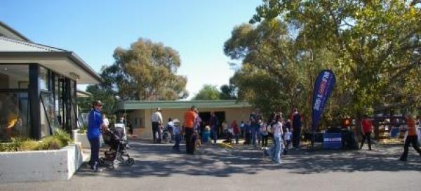 Scenes from Open Day 2009
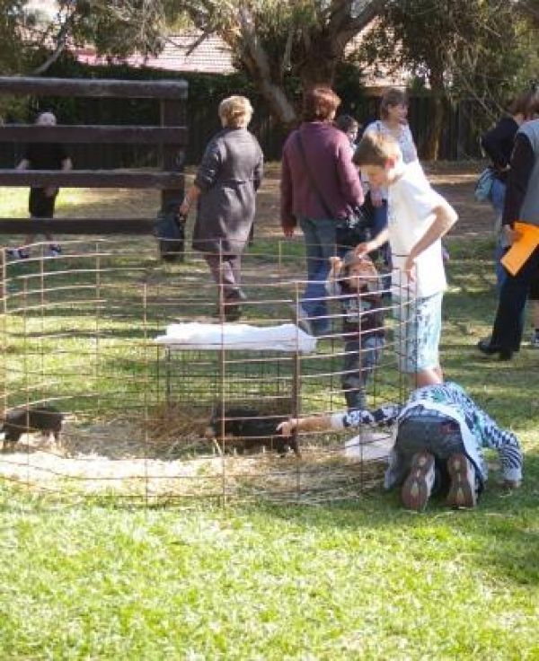 How hard is it to pet a pig?
|
|
|
|
|
|
|
|
|
|
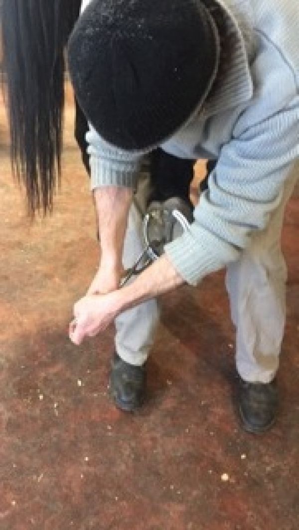
02 Hoof abscesses in horses |
|
|
|
|
Sudden onset lameness in horses very commonly originates from foot pain.
By far and away the most common cause of foot pain is an abscess. Foot abscesses can be so painful your horse may be standing on three legs and very reluctant to move.
Abscesses can originate following a bruise to the sole or heel, white line disease (seedy toe), laminitis, a close nail, or a penetrating injury to the foot. There is very little room for an infection to spread inside the hoof so an infection causes significant pressure on the sensitive tissue causing severe pain and lameness.
Abscesses can occur in any horse, from a Group 1 contender, to a paddock ornament. Both shod and unshod horses, living in stables or paddocks, can develop abscesses at any time of the year.
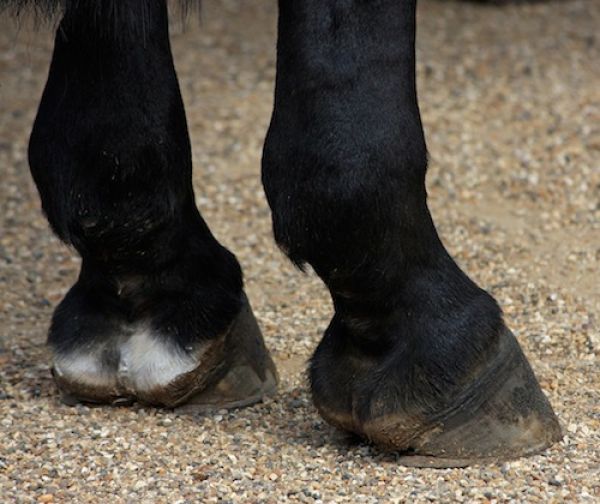
The horse may hold the affected leg up, reluctant to bear weight on the sore foot. The hoof may feel warm to touch and a bounding pulse provides a useful clue that the foot is painful. The digital pulse can be felt on either side of the fetlock at the back of the leg.
Treatment is aimed at allowing the abscess to drain. We can apply hoof testers to the hoof to localise the pain, then carefully explore the hoof in an attempt to make a drainage hole. Digging large holes in the foot searching for the abscess is strongly discouraged. Occasionally abscesses will burst out the heel area or coronary band.
If the abscess cannot be drained in the first instance a poultice bandage can be applied or the foot can be soaked to soften the affected area, thereby allowing the abscess to more easily burst out.
Antibiotics are not generally required for uncomplicated hoof abscesses. We can provide pain relief to make the horse more comfortable allowing a faster recovery and return to work.
The hoof should be kept clean during healing, and care taken to ensure the drainage hole does not clog with dirt or debris until the abscess has fully resolved.
In any case of acutely or severely lame horses call us without delay - 63618388.
|
|
|
|
|
|
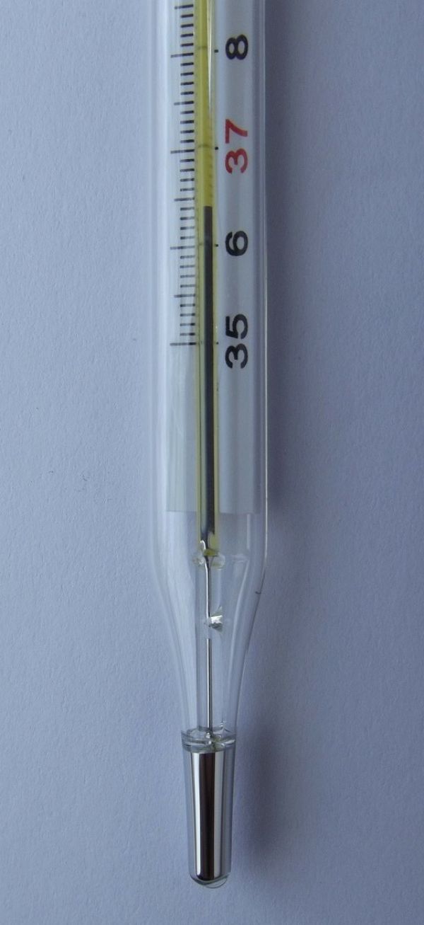
03 Catch Q fever and you may never be the same again |
|
|
|
|
There has been a considerable amount of press regarding Q fever in the last few months. For those of you who missed it, Landline featured this issue in July.
In the past, we have discussed various zoonotic diseases (diseases which can be transmitted from animals to humans). These include leptospirosis and Hendra virus to name a couple. Q-fever is a disease transmitted primarily from cattle, goats and sheep, which can have life long impacts including post Q fever fatigue syndrome. Every year there are hundreds of reported cases of Q fever, and it is likely the disease is under reported.
We do see clinical cases of Coxiella (the bacteria which causes Q fever), particularly in ruminants. Most of the time, an animal infected with Q fever shows no signs, however it can cause animals to go off feed and will also cause late term abortions. Coxiella has also been shown to cause sub-clinical mastitis in dairy cows.
As your vet, we aim to promote a healthy and happy lifestyle shared with your animals. The last thing we want to hear is that one of our clients has caught as disease from the stock on their farm which could have been prevented. If you’re working in the meat and livestock industry, or on any farms in Australia, it is worth considering vaccination.
Some good information is available in the Department of Health's Immunisation Handbook.
|
|
|
|
|
|
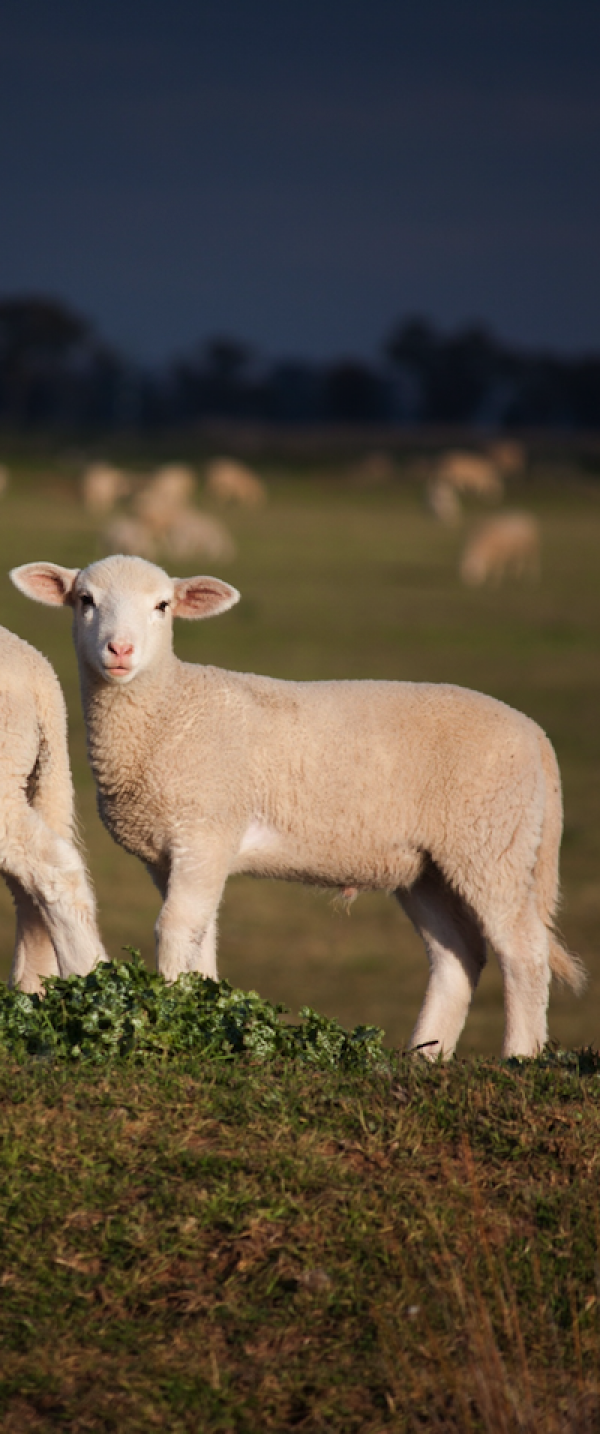
04 The biggest cost to sheep producers |
|
|
|
|
The MLA’s report on diseases affecting the red meat industry put lamb neonatal mortalities right at the top of the list. The production losses associated with neonatal mortalities cost an astounding $442 million dollars to Aussie producers every year.
Lamb mortality is one of the most frustrating aspects of keeping sheep. Working so long to produce a lamb that dies soon after birth is a waste and a profit killer. It is estimated that 20-25% of lambs born never reach weaning (with enormous variation). So what can be done?
Put simply, any management practice that ensures correct nutrition of the pregnant ewe, avoidance of dystocia and adequate lamb nutrition will minimise losses.
Pre-joining
Select ewes intensively for ‘easy-care’ lambing. Although the trait has low heratibility, slow progress does pay off. Buying rams that produce low birth weight lambs is essential to reduce dystocia rates.
During pregnancy
Feed sheep well during pregnancy. There have been multiple attempts to alter feeding during pregnancy to reduce lamb birth weight. It has been met with disappointment. Ewes should be going into 1200kg DM/ha pasture in the four weeks before lambing. Scan the ewes at 80-90 days of pregnancy, and lamb all those carrying multiples separately in paddocks with shelter.
At lambing
The most common causes of lamb mortality are starvation/mismothering, exposure and dystocia. Hypothermia and lamb starvation are closely linked. Young lambs can take quite a lot of cold weather if they are dry, out of winds and drafts and have a belly full of milk. Give the ewes plenty of undisturbed space to lamb and leave them on their birth sites until they want to move away.
Adequate early lamb nutrition
Adequate nutrition of the pregnant ewe and prevention of dystocia influence early lamb nutrition. This ensures the birth of vigorous lambs that receive adequate maternal colostrum.
This is just a short introduction to this topic - if you have an issue or are worried, give us a call!
|
|
|
|
|
|
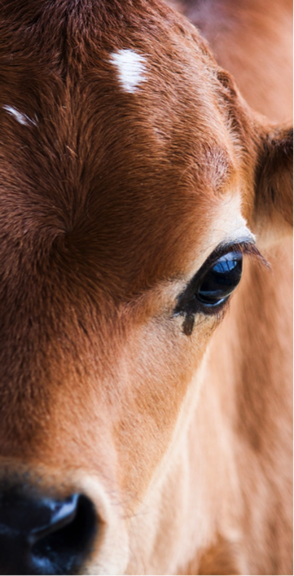
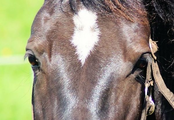
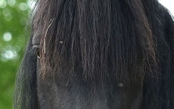
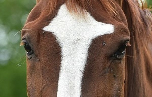
05 What to do about equine eye injuries and ulcers |
|
|
|
|
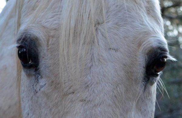
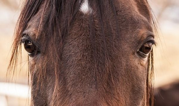
Eye problems in horses are common and can be severe. Severe cases can result in vision loss or requiring removal of a chronically painful eye. Quickly recognising the signs of eye pain is crucial in maximising the chances of effective treatment and speedy recovery.
Horses with painful eyes appear to be squinting as the muscles controlling the eyelids spasm, closing the eye. Discharge or weeping can often occur with damage to the surface of the eye, or conjunctivitis. An ulcer is an area where the protective surface of the tissue is missing.
We all know how painful a sore eye can be, especially if something is stuck in there. Only the most cooperative of horses will let us thoroughly examine the eye without sedation. Grass seeds and other foreign bodies can become lodged deep under the eyelids (including the third eyelid) making them difficult to find and remove.
After sedating the horse we will thoroughly examine the eye and under the eyelids. Staining the eye with a fluorescent dye will allow us to see defects to the surface not otherwise visible. A special tool called an opthalmoscope may be used to see the internal structures of the eye, which are important for vision.
After thoroughly assessing the eye a treatment plan can be begun. Treatment often involves application of ointment or drops containing antibiotics and atropine. Antibiotics are used to prevent or treat any infection that can develop on or below the surface of the eye. Atropine helps to dilate the pupil, aiding treatment and providing pain relief. Some horses will become very difficult to treat and in some cases a treatment tube can be surgically placed to make treatment much easier.
Uncomplicated eye injuries or ulcers often improve quickly with the above treatment. In addition to topical ointments or drops we will often prescribe pain relief. Severe complications are rare with eye injuries but given the importance of the eye to a horse we should be proactive in seeking prompt veterinary attention.
Please contact us if your horse’s eye remains painful after treatment.
|
|
|
|
|
|
|
|
07 BULLCHECK™ - What's involved? |
|
|
Pregnancy rates in cattle herds vary from very good to very bad. Some of the poorest results are achieved when bulls fail and pregnancy rates can drop well below 50% for any given joining period. To help avoid this scenario, BULLCHECK™ was devised to screen bulls prior to a joining to identify issues which are high risk of causing reproductive failure.
|
|
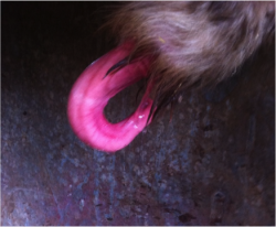
For example, a persistent frenulum (pictured left) is a condition seen, especially in young bulls, which prevents the bull from fully protruding their penis from the sheath; in most cases they will not achieve intromission. A pre-joining BULLCHECK™ will detect this abnormality, preventing a very disappointing joining!
|
What is a BULLCHECK™?
The BULLCHECK™ is a relatively quick and economical procedure ($70-100/bull) for screening bulls prior to sale or use. The Australian Cattle Veterinarians (ACV) have developed world-recognised procedures and standards for conducting BULLCHECKs and for computerising the relevant information.
What does it involve?
Usually, a BULLCHECK™ includes these procedures:
- A general physical examination
- A reproductive examination (including measurement of testicle size, and palpation of the testicles and sex glands).
- Collection and examination of semen (particularly for sperm motility and morphology).
In addition, a serving ability test may be carried out, as may special tests for diseases (e.g. vibriosis or trichomonosis). Although these procedures add predictive value to the BULLCHECK™ and may be indicated in some situations, they are not mandatory.
Following these tests, bulls are placed into one of four categories;
 Satisfactory: All factors assessed were consistent with ACV standards. No risk factors for reduced fertility identified. Satisfactory: All factors assessed were consistent with ACV standards. No risk factors for reduced fertility identified.
 Unsatisfactory: Some factors assessed were not consistent with ACV standards. E.g. Lameness, penile injury or semen morphology defects. Unsatisfactory: Some factors assessed were not consistent with ACV standards. E.g. Lameness, penile injury or semen morphology defects.
 Classification Deferred: Although not all factors met ACV standards, this probably would not preclude using the bull under certain conditions. E.g. Mild post-leg – likely to develop arthritis prematurely. Classification Deferred: Although not all factors met ACV standards, this probably would not preclude using the bull under certain conditions. E.g. Mild post-leg – likely to develop arthritis prematurely.
 Not tested: Factor(s) could not be adequately evaluated. A retest is often recommended. Not tested: Factor(s) could not be adequately evaluated. A retest is often recommended.
In Summary
Where natural breeding bulls are employed, BULLCHECK™ should be an integral management tool for optimising herd fertility, genetics and profitability. If done pre-joining, 1-3 months prior to the joining is ideal.
|
|
|
|
|